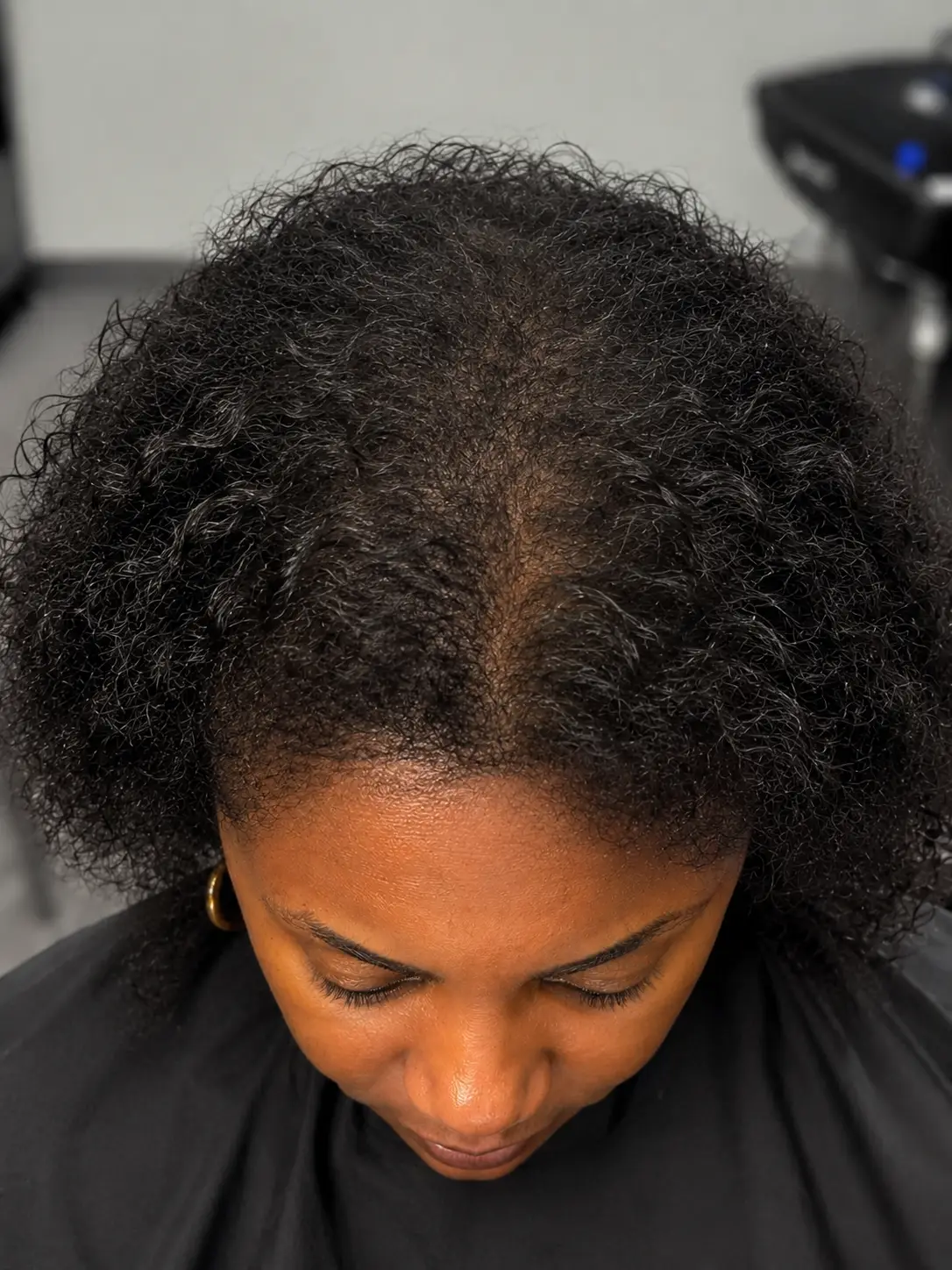
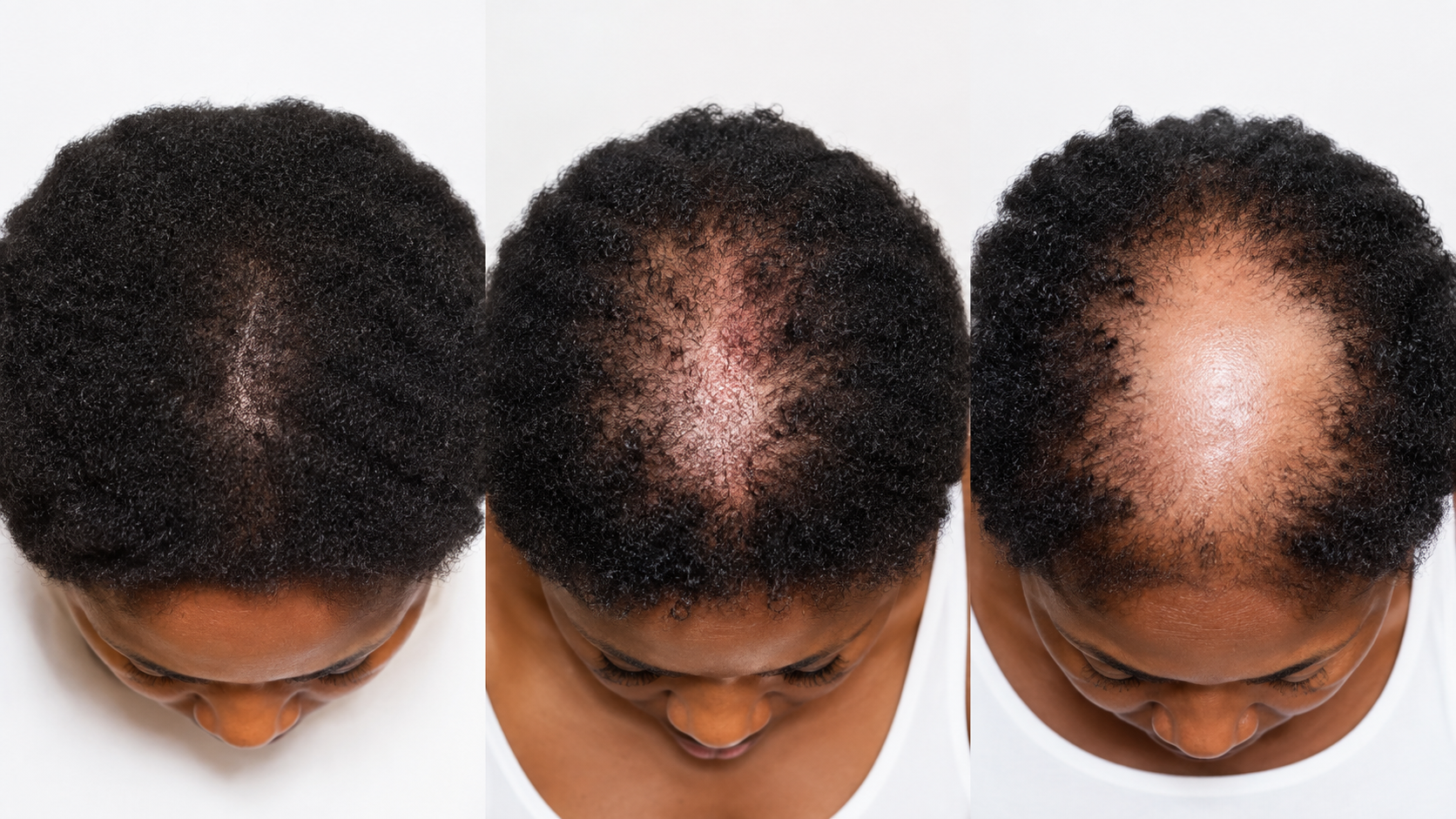
Why staging matters
CCCA is progressive. The earlier it is identified, the more follicles survive. Most women do not learn they have it until thinning is obvious, and by then a meaningful share of the crown has already scarred.
Understanding the stages helps you recognize the early signals while the treatment window is still open. Start with our CCCA condition page for the clinical background.
Stage 1: Subclinical inflammation
No visible thinning. Under trichoscopy, peripilar grey-white halos appear around crown follicles. Mild itching or tenderness may come and go.
This stage is only detectable with magnification. Treatment here can prevent visible hair loss from ever developing.
Stage 2: Early thinning at the crown
Visible thinning starts at the vertex and the part line widens. Trichoscopy shows follicular miniaturization, reduced density per square centimeter, and expanding peripilar halos. Some follicular openings are already gone.
This is the critical treatment window. Most follicles are still viable.
Stage 3: Established scarring with an active border
A clear thin or bald patch sits at the crown with smooth, shiny skin at the center. Trichoscopy shows complete loss of follicular openings in that center, while the border still shows follicles under active attack.
Treatment focuses on halting the border and saving what remains around it.
Stage 4: Advanced scarring
A large area of smooth, scarred scalp extends outward from the crown. Trichoscopy shows widespread absence of follicular openings, sometimes with a residual active border at the edges.
The goal shifts to stopping further spread and maximizing whatever viable follicles remain.
What trichoscopy reveals at each stage
The markers are specific. Peripilar grey-white halos are considered pathognomonic for CCCA. Loss of follicular openings confirms scarring. Pinpoint white dots, the lonely hair sign, and miniaturization patterns fill in the rest of the picture.
None of these are visible to the naked eye or on a standard visual exam. That is the entire argument for imaging before treatment.
Treatment approach by stage
Stages 1 and 2 call for aggressive anti-inflammatory work: PRP, exosomes, laser therapy, and mesotherapy to halt inflammation and preserve every follicle.
Stage 3 shifts to stabilizing the border and regenerating at the margins, often adding microneedling where openings remain. Stage 4 aims at stopping spread, maximizing viable follicles, and pairing clinical care with cosmetic solutions for scarred zones. See scarring alopecia for how CCCA fits the broader category.
What are the stages of CCCA alopecia?
CCCA progresses through four recognizable stages. Stage one is subclinical inflammation, with no visible thinning and peripilar grey-white halos visible only under trichoscopy at 200x magnification. Stage two brings early crown thinning, a widening part, follicular miniaturization, and the first losses of follicular openings, which makes it the most productive treatment window. Stage three shows established scarring at the center of the crown with an active inflammatory border where follicles remain under attack, so treatment concentrates on halting that border. Stage four is advanced scarring across a large area, where the objective becomes stopping further spread. Trichoscopy determines the stage by examining peripilar halos, follicular openings, pinpoint white dots, and miniaturization patterns, none of which are visible on standard examination. Clinical literature consistently reports that outcomes correlate with the stage at which treatment begins, which is why early imaging matters more than any single therapy.
Find out where you are
Book a $99 trichoscopy evaluation with a certified trichologist. 45 minutes. Call (678) 561-4522. We are at 8735 Dunwoody Place, Suite 290-O, Sandy Springs, GA 30350. Related reading: CCCA, the silent hair loss and scarring alopecia in Black hair.