

Condition
Scarring Alopecia Treatment: Save Follicles Before the Damage Is Permanent
Permanent hair loss caused by inflammation that destroys follicles and replaces them with scar tissue.
Map Your Scarring Alopecia with TrichoscopyOur Approach
How We Treat Scarring Alopecia (Cicatricial Alopecia)
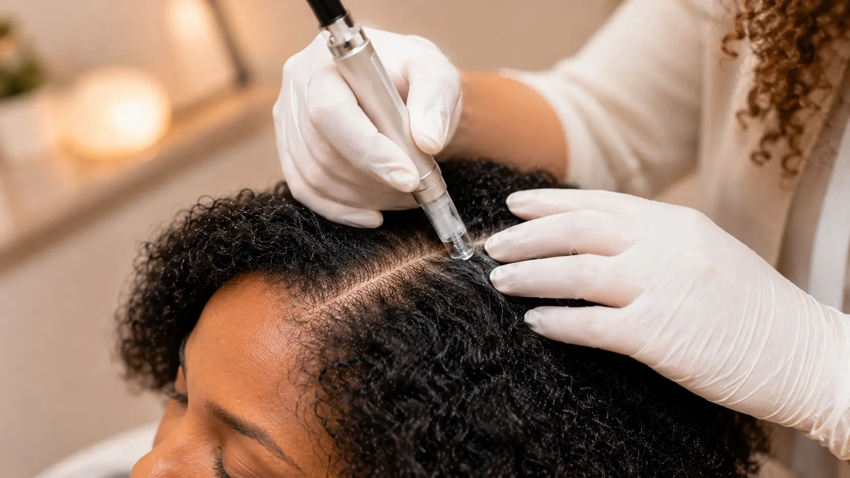
Our team attacks the inflammation to halt progression and protect every viable follicle. Dr. Nina Ross designs each protocol using a combination of PRP, exosome therapy, laser therapy, and anti-inflammatory scalp treatments. Our certified trichologists monitor your scalp with serial trichoscopy imaging to make sure the active border is shrinking. If bloodwork reveals internal inflammatory drivers, our functional medicine team addresses those too.
Explore our treatment methods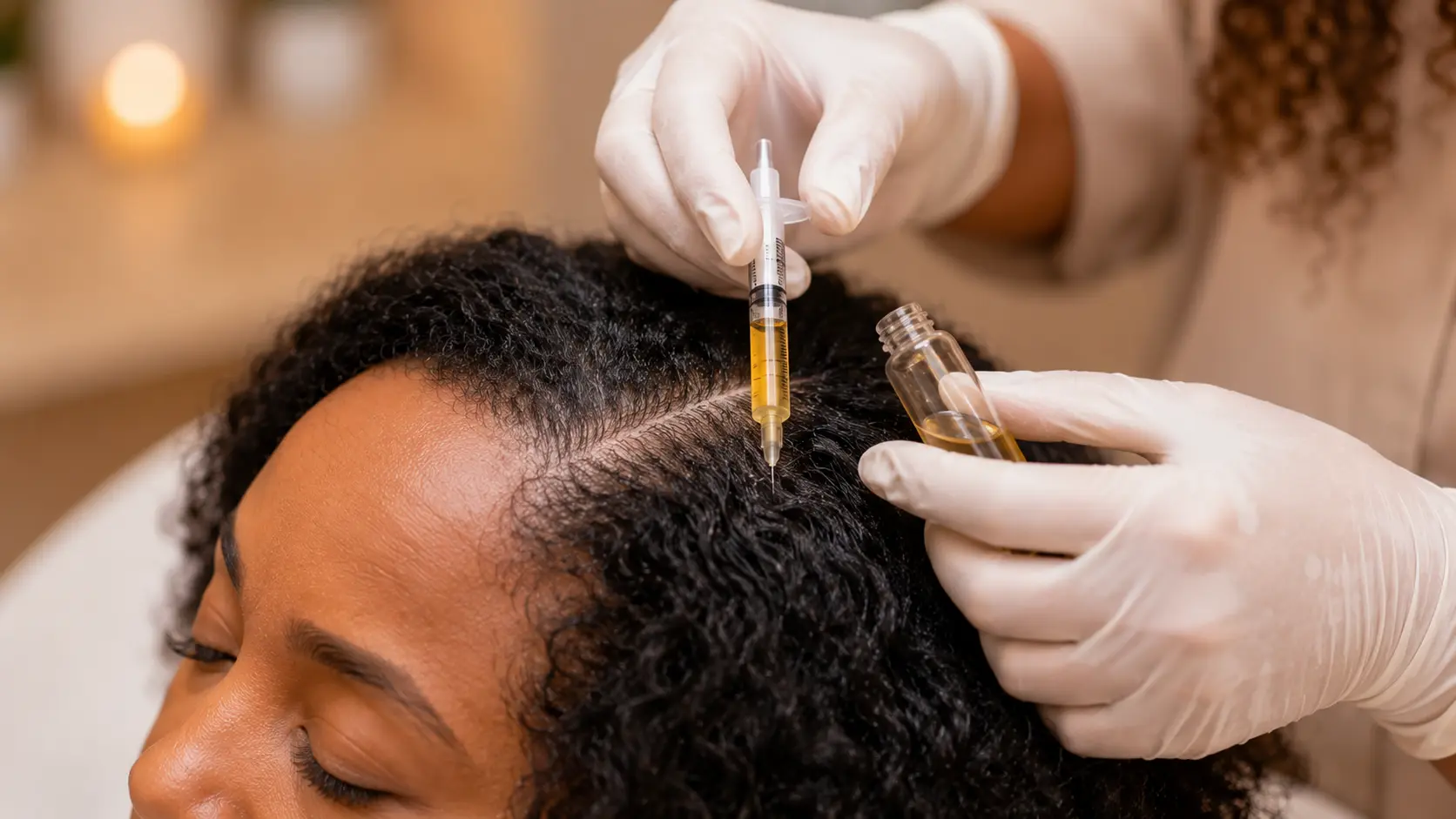


What Scarring Alopecia Is and Why Timing Decides the Outcome
Scarring alopecia, also called cicatricial alopecia, refers to a group of inflammatory conditions that permanently destroy hair follicles and replace them with scar tissue. Major types include CCCA (most common in women of African descent), lichen planopilaris, frontal fibrosing alopecia, and folliculitis decalvans. These conditions share a common pattern: an active inflammatory front that gradually expands, destroying follicles in its path. Once a follicle is replaced by scar tissue, it cannot be recovered. However, follicles at the active border are still alive and responsive to treatment. Trichoscopy at 200x magnification identifies the active border, the scarred zone, and the healthy tissue, enabling precise treatment targeting. At Atlanta Trichology, Dr. Nina Ross (practitioner) designs protocols combining PRP, exosome therapy, laser therapy, and anti-inflammatory treatments. Certified trichologists perform evaluations and track the inflammatory border with serial imaging. The $99 evaluation determines the type and stage. (678) 561-4522.
Trichoscopy at 200x magnification differentiates the sub-types and separates viable follicles from scarred ones. That distinction defines the treatment plan. Atlanta Trichology's Practitioner-Led team identifies the type and stage, then targets therapy at the active inflammation border where follicles can still be rescued. Certified trichologists perform every scan. Dr. Nina Ross, the practitioner and architect of each protocol, reviews the imaging and builds the plan.
Evidence
The Clinical Picture Behind Cicatricial Alopecia
Cicatricial alopecias are defined histologically by destruction of the follicular stem cell bulge and the sebaceous gland, followed by replacement of the follicular unit with fibrous tract. Once that replacement completes, no therapy currently available regrows hair from that site. This is why the clinical objective is halting the advancing border rather than reviving scarred scalp.
The North American Hair Research Society classifies cicatricial alopecias by the dominant inflammatory infiltrate. Lymphocytic types include CCCA, lichen planopilaris, and frontal fibrosing alopecia. Neutrophilic types include folliculitis decalvans and dissecting cellulitis. Identifying the infiltrate pattern guides whether the protocol leans anti-inflammatory, antimicrobial, or both.
Trichoscopic markers are type-specific. Peripilar grey-white halos point toward CCCA. Perifollicular scaling with violaceous erythema points toward lichen planopilaris. A receding band with lone hairs and eyebrow loss points toward frontal fibrosing alopecia. Tufted follicles with pustules point toward folliculitis decalvans. These signs appear at the border before visible loss reaches it.
Studies in the British Journal of Dermatology report that patients treated during the early inflammatory phase show significantly higher rates of stabilization than those who present with established scarring. Time is the single strongest predictor of outcome in this condition group.
Atlanta Trichology documents every visit with comparison trichoscopy so border movement is measured rather than guessed. Dr. Nina Ross reviews the imaging series and adjusts the protocol when the border shows renewed activity.
Investment
Scarring Alopecia Treatment Cost in Atlanta
The scalp evaluation is $99 and runs 45 minutes. A certified trichologist performs 200x trichoscopy, maps the active border, counts viable follicular openings, and documents the baseline for comparison at future visits.
Treatment protocols generally range from $250 to $600 per session depending on the modality combination. Scarring alopecia typically requires 8 to 12 sessions in the first year, followed by maintenance, because suppression of the inflammatory border is an ongoing task rather than a one-time fix.
Packages and payment plans lower the per-session cost for multi-visit protocols. Call (678) 561-4522 to talk through pricing for your stage and sub-type.
Timeline
What the First Year Looks Like
Scarred areas do not regrow. Every gain comes from the border, which is why early scanning changes the ceiling on what is possible.
Common Questions
Scarring Alopecia Questions
Related
You May Also Want To Read

Condition
Traction Alopecia
Hair loss caused by tight hairstyles and chronic tension on the follicle.
Read more
Condition
Alopecia Areata
Autoimmune hair loss that appears as smooth, round patches on the scalp.
Read moreCondition
Central Centrifugal Cicatricial Alopecia (CCCA)
Scarring alopecia beginning at the crown, most common in Black women.
Read moreTreatment
Treatments for Scarring Alopecia (Cicatricial Alopecia)
Your hair is telling you something. Let's find out what.
A 30-minute evaluation. A certified trichologist. A 200x trichoscopy scan of your scalp. And real answers before you leave the building. Your next step is $99 and one appointment away.